Signs and Symptoms
‘Ankle impingement is defined as painful
mechanical limitation of full ankle movement secondary to osseous and/or soft
tissue abnormality’ [13]. This means that the pain that is felt in the back of
the ankle can be due to some bone impinging or squeezing in on things like
muscles, nerves, tendons, ligaments and other structures that are found in the
ankle.
Posterior ankle impingement syndrome (PAIS) can
present with the following symptoms:
- Swelling at the back of the ankle behind the achilles tendon above
the heel. Swelling will become more prominent when the toes are pointed [9,
13]

- Pain on the posterior (back) of the ankle which is exacerbated by
plantarflexion (toes pointed) and dorsiflexion (pulling foot up towards
shin) [8, 13]
- Less than normal movement for plantarflexion and dorsiflexion [9, 11, 13]
 |  |
Differential Diagnosis
If these symptoms are present then there is a
chance that you may have PAIS [10, 11, 13, 16]. However just because you have the
symptoms of PAIS does not necessarily mean you have it. There are several other
conditions that have similar presentations as PAIS:
- Osteochondral injury [4 ,10 , 13] –
www.footandankle.mdmercy.com/conditions/ankle_injury/osteochondral.html
- Mechanical instability [9, 10, 13] –
- Peritendonitis [9, 10, 13] –
www.wrongdiagnosis.com/medical/peritendinitis.htm
- Flexor
hallucis longus tenosynovitis [9, 10, 13] –
www.arthritis-treatment-and-relief.com/flexor-hallucis-longus-tendon-problems.html
- Peroneal
tenosynovitis and tendonitis [5, 10, 13] –
www.aidmyplantar.com/peroneal-tendonitis.php
- Intra-articular
loose bodies [9, 10, 13] -
- Ankle
synovitis [9, 10, 13] -
www.allcures.com/shared/conditions.asp?id=303
The website under each condition will give some more information as to
what each is and what they look like.
Diagnostic Tests
The following
video demonstrates what a physiotherapist or doctor may do to see if a patient
has the signs or symptoms of a PAIS. [Youtube.com]
In the video you’ll notice that the therapist is palpating (feeling or touching) at the back of the ankle to see if this is painful. Also he is checking the how far the ankle can move up and down (toe pointing and lifting toes) while asking the patient if the movement hurts.
Diagnostic
Investigations
To be completely sure of what is causing the symptoms (pain, swelling
and less movement); several tests can be performed. These tests include:
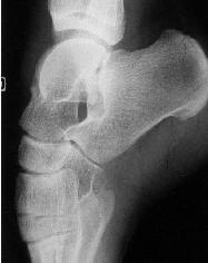
- Conventional radiograph (x-ray) - Conventional radiographs can
demonstrate any bony abnormalities such as an os trigonum or a Steida
process [13]. An os trigonum is a piece of bone that has come loose from the
highest bone in the ankle (the talus) and can sometimes pinch soft tissue like
muscles, ligaments or tendons [9]. A Steida process is a large bony bump
or tubercle that also comes off the talus (highest bone in the ankle) [13].
This bony bump can also cause impingement of muscles, tendons or ligaments
among other things. X-rays however can sometimes fail to detect stress
fractures (small cracks that appear in the bone) [13]; this means that
while a patient may still be experiencing pain, the x-ray image would show
no problem when in actual fact there is something wrong. Below is an x-ray image of the ankle
- CT scan – CT scans are much like x-rays but can demonstrate a much more detailed image of bony structures, enabling them to detect tiny cracks in the bones (stress fractures) [6]. However like x-rays a CT cannot show a good image of soft tissues like muscle [2].
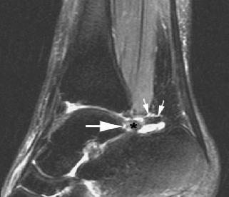
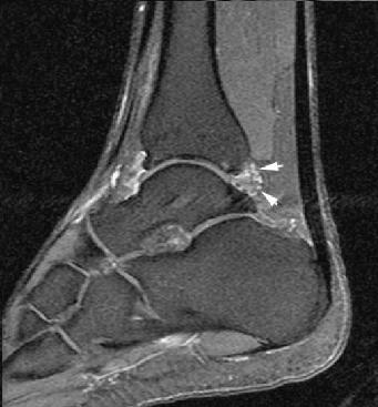
- MRI – MRI is
by far the best test for detecting PAIS [2, 6, 18]. This is
because it detects water, so can show a clear image of soft tissue as well
as bone. It is also useful in assessing any surrounding structures that
may be involved in causing pain [2, 4, 12, 14] (pictures below). This aids physiotherapists
in coming up with the best possible treatment plans, or surgical plan for
doctors [13]. MRI also has the added benefit of not exposing patients to
radiation.
 |  |